My knee is locked!

The knee joint is designed to bend up and down and rotate to a certain degree. If something gets caught inside the joint, it blocks the movement and the knee gets lock.  The locked knee is most commonly as a result of a meniscus tear. Other causes of a locked knee are due to cartilage and bony fragments, ligament injury, loose tissue fragment and patella (knee cap) dislocation. In this article, I would like to focus on the meniscus injury as it is the most common cause of locked knee.
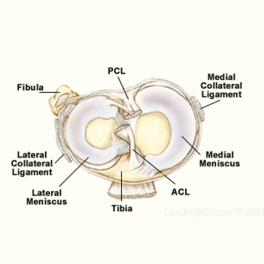
The meniscus is a rubbery, C-shaped cartilage in between the femur (thighbone) and tibia (shinbone). There are two menisci in each knee joint, the inner medial meniscus and the outer lateral meniscus. The outer edges are fairly thick while the inner surfaces are thin. The meniscus provides smooth knee motion, stability and contributes to a healthy knee through its shock absorber effect. During the various phases of the walking cycle, forces shift from one meniscus to the other, and forces on the knee can increase to 2 – 4 times body weight. While running, these forces on the knee increase up to 6 – 8 times body  weight. There are even higher forces when landing from a jump.

The meniscus can be damaged or torn during activities that cause direct hit or pressure from a forced twist. For example, taking a hard tackle on the football field or a sudden turn on the futsal or badminton court can result in a meniscus tear. In elderly, as the meniscus ages, it weakens and become less elastic. Hence, even a minor event such as getting up too quickly from a squatting position or a twisted knee during walking on uneven surface can also tear the meniscus. Apart from locking, meniscus tear can produce symptoms of pain, swelling and imbalanced. It is common for a meniscus tear to occur concomitantly with a knee ligaments or chondral injuries.

Diagnosis is confirmed through a detailed history, physical examination, special test and imaging studies such as the MRI (Magnetic Resonance Image). In competent hands, arthroscopy is the best tool for meniscal tear diagnosis, with sensitivity, specificity, and accuracy approaching 100%. Being both therapeutic and diagnostic, it offers the option of immediate treatment of most disorders.

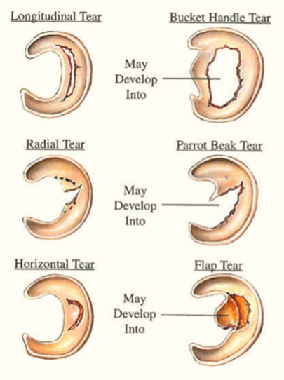
Meniscal tears come in many size and patterns which influences the decision on treatment. Examples of the various tear patterns are: longitudinal, bucket-handle, parrot beak, radial, displaced flap and horizontal. The bucket-handle tear is the most common meniscus tear which cause locked knee and it requires early surgery.

In the 1960s and 1970s, it was common to remove a damaged meniscus entirely. This frequently led to early degenerative arthritis in many patients. Removing the entire medial meniscus can lead to a bow-legged deformity and medial joint arthritis while removing the entire lateral meniscus can cause a knock-kneed deformity and lateral joint arthritis.

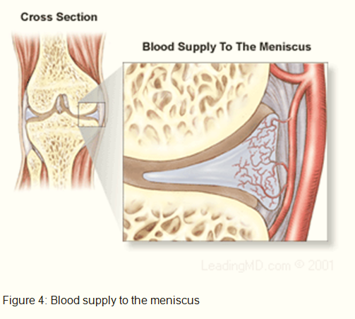
Currently, treatment decision is based on the patient’s activity level, age, location, size and type of tear, duration of injury and the presence of associated knee injuries. In the acute phase, the injured knee is put on rest, ice and compression bandage. Together with anti-inflammatory medications, these can reduce pain and swelling. Many small meniscal tears will heal without surgical treatment. Partial tears, degenerative tears, and stable tears may be observed for 2 – 3 months. However, a large meniscus tears causing locked knee or with a concomitant anterior cruciate ligament injury are indicated for early surgical treatment. In these cases, a delay in treatment may convert a repairable meniscus tear into a non-repairable tear and subsequent further damage to the knee joint. If surgery is recommended, it is done through an arthroscopy. The procedure chosen is usually dependent on the location, size and type of meniscal tear. The range of surgical treatment includes; Trephination/Abrasion Technique, Meniscal Repair, Partial Resection, Complete Resection and Meniscal Replacement. Trephination/Abrasion Technique is used for stable tears located on the periphery where there's a good blood supply. Multiple holes or shavings are made in the torn part of the meniscus to promote bleeding, which enhances the healing process.

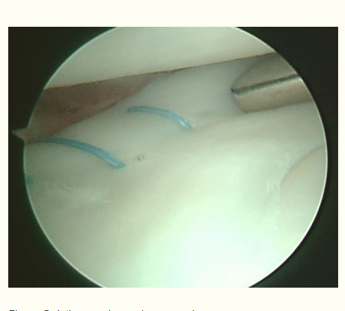
Meniscal Repairs are performed on tears near the outer 1/3 of the meniscus where a good blood supply exists, or on large tears that would require a near-total resection. The torn portion of the meniscus is repaired by using either sutures or absorbable fixation devices. These devices include arrows, barbs, staples, or tacks that join the torn edges of the meniscus so they can heal.

Partial Resection is for tears located in the inner 2/3 of the meniscus where there is no blood supply. The goal is to stabilize the rim of the meniscus by removing as little of the inner meniscus as possible. Only the torn part of the meniscus is removed. If the meniscus remains mostly intact with only the inner portion removed, the patient usually does well and does not develop early arthritis.

Complete Resection of the damaged meniscus is performed if absolutely necessary. Removal of the entire meniscus frequently leads to the development of arthritis. To prevent this, meniscus replacement surgery is done following a complete meniscus resection. The replacement can be done either with a collagen meniscus implant or a meniscal transplant from a donor.

Post-operatively, brace and crutches may be needed. Recovery time depends on number of factors, including the severity of the meniscus tear. Full recovery from surgery may take 4 to 12 weeks, depending on the type of procedure performed. Physiotherapy is needed for a good functional outcome.

To lower the risks of a meniscus injury requires regular exercises to keep the leg muscles strong, maintain flexibility, warm up before sports activities, adequate rest in between workout to prevent muscle fatigue, wear shoes with adequate support and fit correctly and never abruptly increase the intensity of workout.